
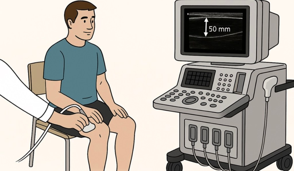
A new publication by Thomas G. Balshaw, Mark P. Funnell, Emmet J. McDermott, Thomas M. Maden-Wilkinson, Lewis J. James and Jonathan P. Folland has examined the accuracy of ultrasound muscle thickness measurements for assessing resistance training-induced muscle growth, with findings that could have important implications for researchers and practitioners working in sport and exercise science.
Summary
Research has supported the observation that individuals with larger muscles tend to be stronger than those with smaller muscles, with resistance training (RT) induced changes in muscle size (i.e., muscle volume, cross-sectional area and thickness) strongly correlated with changes in muscle strength. Ultrasonography is one of the most common approaches used to quantify the changes in muscle size following RT, using muscle thickness (MTUS) measurements, a 2-dimensional measure taken at a single or multiple sites on the muscle which is assumed to be representative of the changes occurring across the muscle. However, despite the widespread use of MTUS measurements to quantify RT induced muscle growth, the gold standard measurement of muscle size remains magnetic resonance imaging (MRI) through 2-dimensional anatomical cross-sectional area (ACSAMRI) and 3-dimensional muscle volume (CSA measures taken along the length of the muscle – VOLMRI) may provide a greater index criterion for the observed changes. Therefore, to better understand how sensitive MTUS measures are relative to the gold standard MRI (ACSAMRI and VOLMRI) for quantifying RT induced muscle growth. We recruited 39 young healthy individuals who performed 15 weeks of supervised lower-body progressive overload RT (knee-extension, knee flexion and leg press; x3 times per week). Pre and post RT measures of MTUS vs ACSAMRI and VOLMRI for the quadricep muscles were performed and manually analysed. It was found that MTUS measurements of the quadricep muscles following RT underestimated the magnitude of overall quadricep muscle growth by 7.3-7.8% relative to measurements ACSAMRI and VOLMRI, mainly due to underestimation of the changes in vastus lateralis and vastus medialis muscles. Therefore, it can be concluded that MTUS measurements underestimate muscle growth following 15 weeks of RT when compared to a valid measure of muscle growth in MRI; and consequently, practitioners should exercise caution when interpretating MTUS derived measures of muscle growth in response to RT.
Citation
Balshaw, T. G., Funnell, M. P., McDermott, E. J., Maden-Wilkinson, T. M., James, L. J., & Folland, J. P. (2026). Ultrasound muscle thickness is a poor index of criterion magnetic resonance imaging measures of resistance training-induced muscle growth. Medicine & Science in Sports & Exercise.
Discover more from PESS News & Information
Subscribe to get the latest posts sent to your email.
